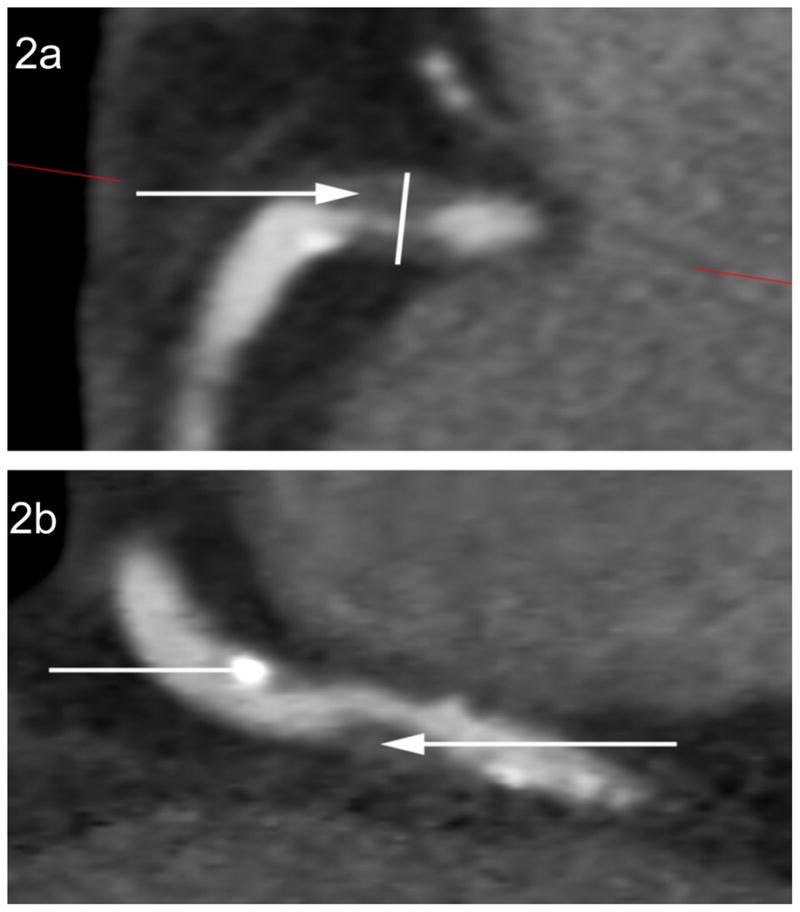
Figure 2.

The assessment of HRPFs. Fig. 1a demonstrates the presence of three high-risk plaque features within the proximal right coronary artery: a >70% stenosis, LAP (closed arrow) and PR at the site of non-calcified plaque (open arrow). Fig. 2b demonstrates the presence of a proximal partially calcified plaque within the right coronary artery (open arrow) and a more distal non-calcified plaque with a LAP component (closed arrow).
HRPFs – high-risk plaque features; LAP – low attenuation plaque; PR – positive remodelling.
