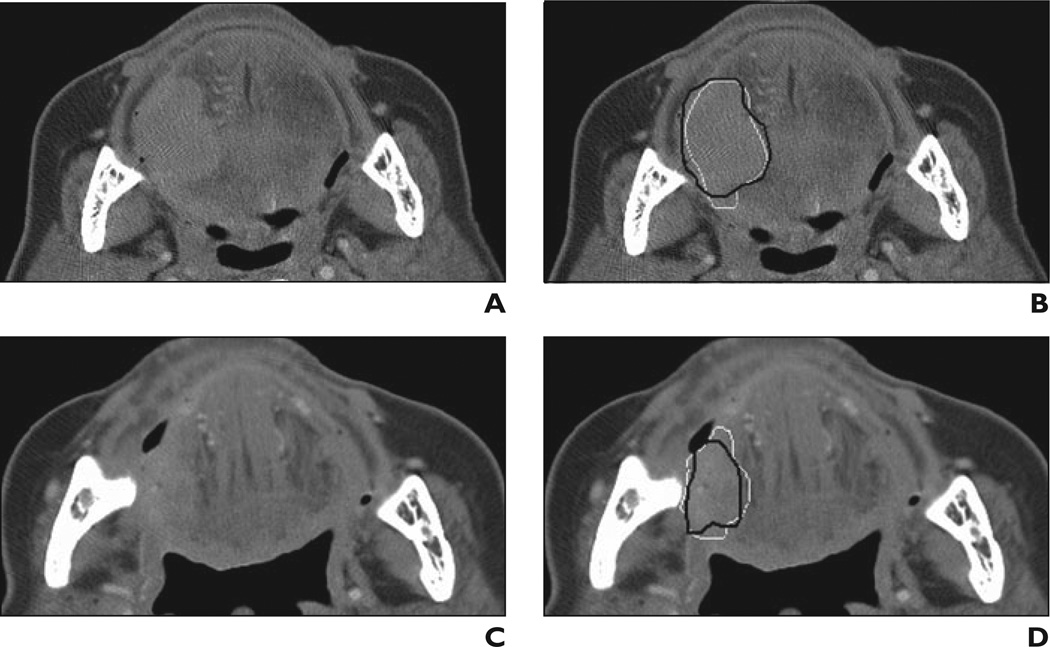
Fig 1. 81-year-old woman with tongue base carcinoma. This lesion is example of subtle lesion (difficulty rating = 4) in data set.
A, Axial CT scan obtained before treatment.
B, Reference standard (i.e., hand-drawn) segmentation (black contour) and automatic segmentation (white contour) are shown superimposed on pretreatment scan.
C, Axial CT scan obtained after treatment.
D, Reference standard segmentation (black contour) and automatic segmentation (white contour) are shown superimposed on posttreatment scan. Lesion is shown on best slice marked by radiologist for each scan.

