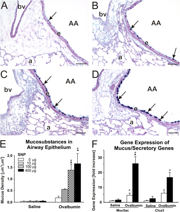
Figure 5.

Airway epithelial mucus production. Increase in airway epithelial mucus, as a feature of allergic airway disease, was analyzed on lung tissue at the 5th generation of the intraepithelial AB/PAS stained mucosubstances (arrows) in SNP/OVA-mice with increasing SNP exposure dose are shown in figure A (0 μg), B (10 μg), C (100 μg) and D (400 μg); AA = axial airway, e = airway epithelium, a = alveoli, bv = blood vessel. Morphometric measurement of intraepithelial AB/PAS mucosubstances are further shown in E and changes in Muc5ac and Clca3 gene expression in F. •: Significant changes (p < 0.05) when compared to non-SNP exposed animals, *: significant changes when compared to non-allergic controls.
