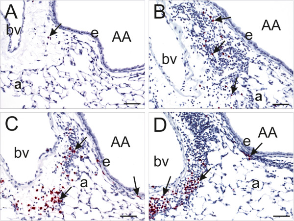
Figure 6.

Immunohistochemistry of airway-associated eosinophils. Light photomicrographs of peri-bronchiolar and –vascular interstitium surrounding the proximal axial airway (AA) at generation 5. Tissues were immunohistochemically stained for eosinophils (murine-specific anti-major basic protein antibody; red chromagen; arrows) and counterstained with hematoxylin. OVA-induced inflammatory cell infiltrate composed of eosinophils and mononuclear cells (lymphocytes and plasma cells) is dose-dependently enhanced by SNP. Figures A-D are taken from OVA-treated mice that were co-exposed to 0 (saline control), 10, 100 and 400 μg SNP, respectively. bv: blood vessel; e: airway epithelium; a: alveolus; Scale bars = 50 μm.
