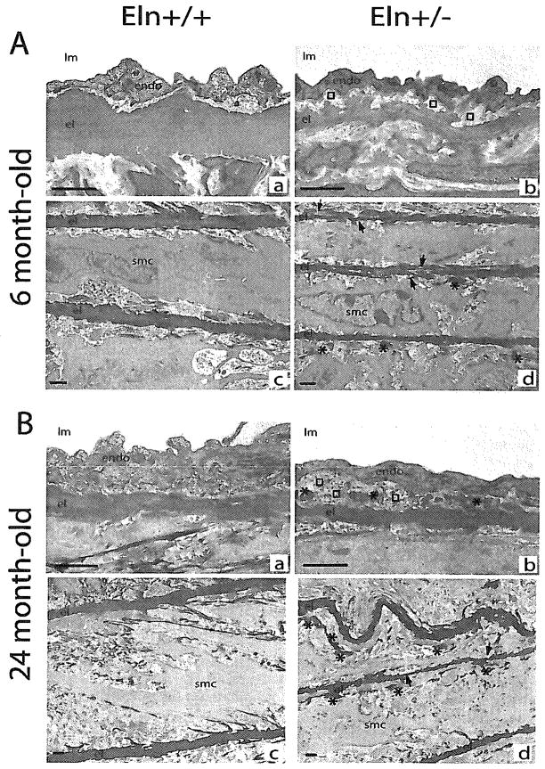
FIG. 2.
Transmission electron microscopy images of the ascending aorta in mice aged of 6 (A) and 24 months (B). (a,c) Eln+/+; (b,d) Eln+/−; (a,b) intima; (c,d) media. Arrows, disruptions of the elastic lamella; asterisks, elastin deposit in margin of elastic lamellas; squares, subendothelial accumulation of extracellular matrix; endo, endothelium; el, elastic lamella; lm: lumen; smc, smooth muscle cell. Bar size: 1 μm.

