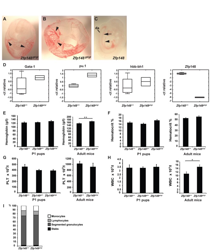
Figure 1. Primitive and embryonic hematopoiesis in Zfp148-deficient embryos and mice.
(A–C) Blood filled vessels in E9.5 Zfp148 gt/gt embryo and yolk sac. (A) Embryo in yolk sac. (B) Primitive vascular plexa in Zfp148 gt/gt yolk sac (arrowheads). (C) Apparent presence of major blood vessels in Zfp148 gt/gt embryo proper (da: dorsal aorta; arrow indicating posterior cardinal vein; arrowhead indicating anterior cardinal vein). (D) Real-time RT-PCR showing relative levels of biomarkers of hematopoietic differentiation. The edges of the boxes show the 25th and 75th percentiles, the central mark is the median, and the whiskers extend to the most extreme data points. (E–H) Blood parameters in peripheral blood from postnatal day 1 pups (P1, left graph) and adult mice (right graph; ages 2-12 months). (E) Hemoglobin, (F) hematocrit and (G) number of platelets in P1 pups; wild type (n = 21), Zfp148 gt/+ (n = 36) and Zfp148 gt/gt (n = 6-7) and adult mice; wild type (n = 19) and Zfp148 gt/gt (n = 15). (H) White blood cells in P1 pups; wild type (n = 7), Zfp148 gt/+ (n = 32) and Zfp148 gt/gt (n = 6). Adult mice; wild type (n = 19) and Zfp148 gt/gt (n = 13). (I) Differential count of blood smears from P1 mice. Percentage of leukocyte populations; monocytes, lymphocytes, segmented granulocytes and stabs in wild type (n = 18), Zfp148 gt/+ (n = 15) and Zfp148 gt/gt (n = 11). Values are expressed as mean ± SEM. Significance levels: *P<0.05, **P<0.01.

