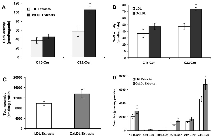
Figure 5. Effect of lipid extracts from LDL and OxLDL on CerS activity.
(A) Oxidation of LDL was performed as described under “Experimental Procedure”. RAW 264.7 cells were stimulated with lipid extracts from native LDL and OxLDL (50 µg protein/mL respectively) for 24 h. CerS activity in cell homogenates was measured using C16-CoA and C22-CoA substrates. Results are means ± S.D., *p < 0.05, of a typical experiment repeated four times with similar results. (B) Cells were treated with intact LDL and OxLDL (50 µg protein/mL respectively) as described above for 24 h. CerS activity in cell homogenates was measured using C16-CoA and C22-CoA substrates. Results are means ± S.D. *p < 0.05, of a typical experiment repeated four times with similar results. (C) Cells were treated as described above and lipids were extracted and analyzed for ceramide content as described under “Experimental Procedure”. No probability values are given for total ceramide levels because these levels are the sum of ceramide species with different acyl chain lengths. (D) Ceramide species were analyzed after treatment with lipid extracts from native LDL and OxLDL as described earlier. The data are means ± S.E., *p < 0.05, n = 4.

