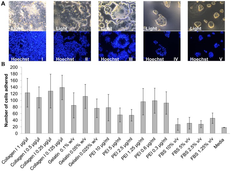
Figure 3. Adhesion studies in adult human cardiomyocytes.
(a) Light and Fluorescent microscope images of adult human cardiomyocytes seeded onto different coatings, took after overnight incubation. (I) Light and Hoechst image for collagen 1 coating (0.125 µg/µl), (II) Light and Hoechst image for gelatine coating (0.05% w/v), (III) Light and Hoechst image for polyethyleneimine coating (0.6 µg/ml), (IV) Light and Hoechst image for foetal bovine serum coating (1.25%), and (V) Light and Hoechst image for no coating (b) adult human cardiomyocytes adhered per view as indicated, and scored using manual counting using ImageJ software, post 1× PBS wash after overnight incubation in the corresponding coatings. Data shown are the media ± SD. n = 4 for all the conditions.

