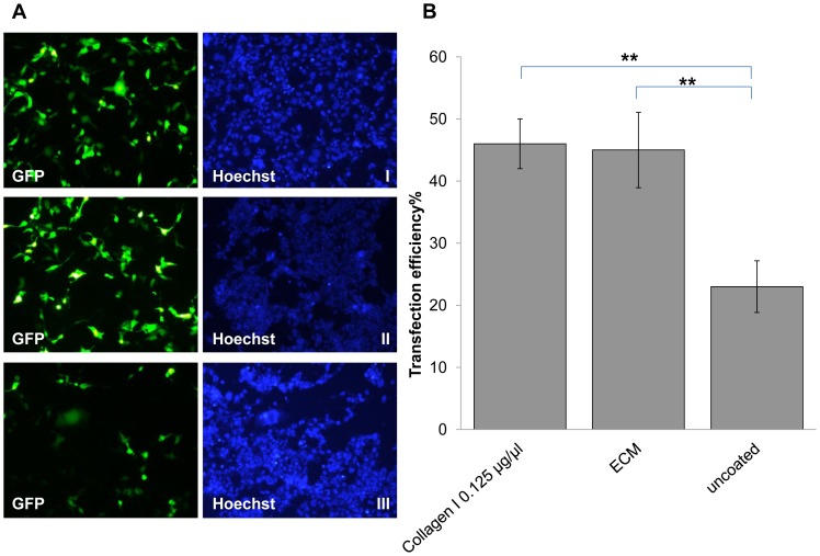
Figure 4. Comparison of oscillating magnet array-based nanomagnetic transfection between adult human cardiomyocytes seeded onto rat tail collagen 1 coated and ECM coated plates.
(a) Fluorescent microscope images of adult human cardiomyocytes transfected with pEGFP-N1 representing each conditions, took 48 hrs post transfection. (I) GFP fluorescence and Hoechst image for 2 Hz 0.2 mm in collagen 1 coated (0.125 µg/µl) plate, (II) GFP fluorescence and Hoechst image for 2 Hz 0.2 mm in ECM coated plate, and (III) GFP fluorescence and Hoechst image for 2 Hz 0.2 mm in un-coated plate (b) adult human cardiomyocytes were transfected with pEGFP-N1 as indicated, and scored using manual counting using Image J software, 48 h after transfection. Data shown are the media ± SD. n = 3 for all the conditions. (**p<0.01- Statistically significant).

