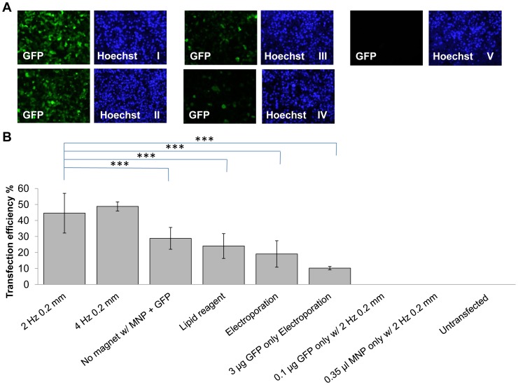
Figure 5. Comparison of oscillating magnet array-based nanomagnetic transfection over lipid based transfection and electroporation in adult human cardiomyocytes.
(a) Fluorescent microscope images of adult human cardiomyocytes transfected with pEGFP-N1 representing each conditions, took 48 h post transfection. (I) GFP fluorescence and Hoechst image for oscillating magnet array-based nanomagnetic transfection performed at 2 Hz 0.2 mm, (II) GFP fluorescence and Hoechst image for oscillating magnet array-based nanomagnetic transfection performed at 4 Hz 0.2 mm (III) GFP fluorescence and Hoechst image for lipid based transfection reagent, (IV) GFP fluorescence and Hoechst image for electroporation and (V) GFP fluorescence and Hoechst image for untransfected (b) adult human cardiomyocytes were transfected with pEGFP-N1 as indicated, and scored using manual counting using Image J software, 48 h after transfection. Data shown are the media ± SD. n = 18 for oscillating magnet array-based nanomagnetic transfection at 2 Hz 0.2 mm. n = 12 for lipid based transfection reagent. n = 6 for oscillating magnet array-based nanomagnetic transfection at 4 Hz 0.2 mm, electroporation and pEGFP-N1 only electroporation conditions. n = 9 for no magnet. n = 3 for pEGFP-N1 only, Neuromag only, and un-transfected conditions. (***p<0.001- Statistically significant).

