
Figure 5.
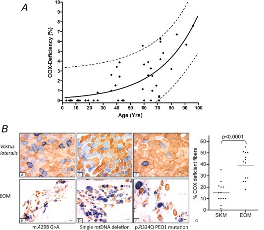
Vulnerability of extraocular muscles to mtDNA mutation accumulation. (A) The data shown in this panel highlight the distribution of COX-deficient extraocular muscle fibres with age in a series of 46 normal human subjects (age range 3–96 years). The simulated curve models an exponential increase in COX deficiency with normal aging (r2 = 0.5753) whilst the dotted lines accentuate the predicted upper and lower 95% confidence range within this group. Data are reproduced from Yu-Wai-Man et al. (2010a) with permission. (B) Enhanced mitochondrial histochemical abnormalities are evident in extraocular muscle compared to skeletal muscle (quadriceps) biopsies in patients with mitochondrial CPEO. Illustrated is sequential COX/SDH histochemistry on paired muscle samples from patients with a mitochondrial tRNA point mutation (m.4298G>A) (a and b), a single, large-scale 5.0 kb mtDNA deletion (c and d) and multiple mtDNA deletions secondary to a p.R334Q PEO1 (Twinkle) gene mutation (e and f). A comparison of the percentage of COX-deficient fibres between skeletal muscle and extraocular muscles in a panel of 13 mitochondrial CPEO patients reveals a significantly higher percentage of COX-deficient fibres in extraocular muscle compared to skeletal muscle (p < 0.0001) (g). Data are reproduced from Greaves et al. (2010) with permission.