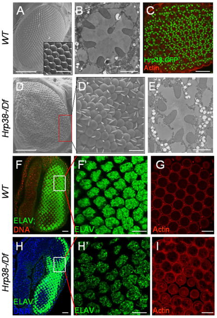
Figure 1. The hrp38 gene is required for normal pattern formation of the fly eye.
(A) Scanning electron microscopy (SEM) image of wild-type eye. Insert shows magnified view of the regular organization of ommatidia and bristles. (B) Tangential sections of wild-type adult retinae showing seven photoreceptor cells in ommatidia. (C) Confocal section of the pupal eye (44h AP) of the Hrp38:GFP strain. F-actin is enriched in rhabodomere. (D,D′) SEM image of the hrp38 hemizygous eye (hrp38d05172/Df) showing the disrupted pattern of ommatidia and bristles. (D′) shows magnified view of boxed area in (D). (E) Tangential sections of the hrp38 hemizygous retinae showing that one photoreceptor cell is missing in an ommatidium. (F) and (F′) The eye imaginal disc of the wild-type fly stained with Elav showing the regular array of the specified photoreceptor cells. The nuclear DNA was stained with Draq5 (red) in (F). (F′) shows magnified view of boxed area in (F). (G) Confocal section of the pupal eye (44h AP) of the wild-type strain stained for F-actin. (H,H′) The eye imaginal disc of the hrp38 hemizygous fly stained with Elav showing the organization of the specified photoreceptor cells. The nuclear DNA in (H) was stained with Draq5 (blue). (H′) shows a disrupted pattern of the photoreceptor cells magnified in the boxed area in (H). (I) Confocal section of the pupal eye (44h AP) of the hrp38 hemizygous fly stained for F-actin, showing the disrupted ommatidial lattice. Scale bars: 100 μm (A, D); 10μm (A insert, C, D′, F, F′,G, H, H′ and I); 2 μm (B, E).

