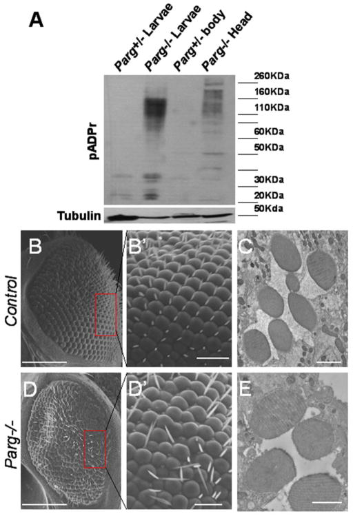
Figure 3. The Parg gene is required for normal pattern formation of the fly eye.
(A) Western blotting analysis of pADPr in the wild-type and Parg mutant eye. Accumulation of pADPr was shown in the adult head, including the Parg mutant eye (Parg−/− head), but not in the separated body (Parg+/− body) dissected from the adult fly (Parg 27.1, FRT19A/FRT19A,GMR-hid; ey-Gal4,UAS-FLP). Parg−/− and Parg+/− larvae were used as the positive and negative controls, respectively. (B,B′) SEM image of the control eye clones (FRT19A/FRT19A,GMR-hid; ey-Gal4,UAS-FLP), showing the regular array of ommatidia and bristles. (B′) shows magnified view of boxed area in (B). (C) Tangential sections of the adult retinae of the control fly (FRT19A/FRT19A,GMR-hid; ey-Gal4,UAS-FLP), showing seven photoreceptor cells in an ommatidium. (D,D′) SEM image of the Parg−/− eye clones (Parg27.1, FRT19A/FRT19A,GMR-hid; ey-Gal4,UAS-FLP), showing the disrupted organization of ommatidia and bristles. (D′) shows magnified view of boxed area in (D). (E) Tangential sections of the adult retinae of the Parg−/− eye clones, showing missing photoreceptor cells in an ommatidium. Scale bars; 100 μm (B, D); 10μm (B′, D′); 2 μm (C, E).

