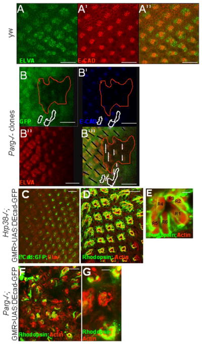
Figure 4. Hrp38 poly(ADP-ribosyl)ation controls DE-cadherin expression in the eye.

(A,A′,A″) DE-cadherin expression pattern in the third-instar eye discs of the wild-type fly (y,w). (B,B′,B″,B‴) Decreased DE-cadherin expression in the Parg mutant clones in the third-instar eye imaginal discs {Parg27.1, P{FRT(whs)101/Ubi-GFP,P{FRT(whshs)101; P{GAL4-ey.H}SS5, P{UAS-FLP1.D}JD2/+}. The Parg mutant clones are GFP-negative and circled in (B,B″,B‴). The black lines indicate the orientation of the wild-type ommatidia in (B‴). The dashed white lines indicate the orientation of the mutant ommatidia in (B‴). The photoreceptor precursor cells were labeled with anti-ELAV antibody in (A) and (B″). (C,D,E) Rescue of the rough-eye phenotype by expression of DE-cadherin:GFP transgene in the Hrp38 mutant background (UAS:DEF/longGMR:Gal4; hrp38do5172/Df). (C) The regular array of the photoreceptor cells labeled with anti-ELAV antibody with DE-cadherin:GFP expression induced by longGMR:Gal4 driver in the third-instar eye disc. (D) The regular ommatidia structure in the adult eye of the rescued fly visualized by confocal imaging. (E) Confocal image of an ommatidium from (D) with the normal seven photoreceptor cells. (F,G) Failed rescue of the Parg mutant phenotype with ubiquitous expression of DE-cadherin (Parg27.1, FRT19A/FRT19A,GMR-hid; Tub-DE-cadherin/eye-Gal4,UAS-FLP). (F) The irregular ommatidia structure in the Parg mutant eye. (G) Reduced photoreceptor cell numbers in the Parg mutant eye. Adult photoreceptor cells in (D,E,F,G) were labeled with anti-Rhodopsin antibody and F-actin. Scale bars: 10 μm (A, A′, A″, B, B′,B″, B‴, C); 100 μm (D, F); 5 μm (E, G).
