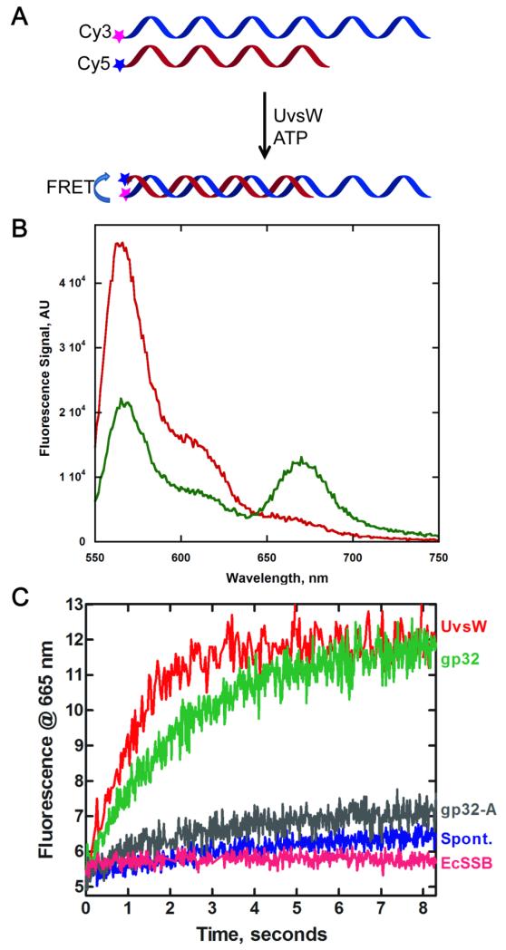
Fig 3.
Effect of ssDNA binding proteins on the kinetics of the annealing activity of UvsW protein. (A) Schematic representation of a FRET-based assay to monitor UvsW mediated annealing of complementary ssDNA in the presence of ATP. The annealing reaction brings the Cy3 and Cy5 pair together that causes a FRET between the dyes and the emission from Cy5 was used to monitor the annealing reaction of the complementary DNA strands. (B) Steady state fluorescence measurements of the UvsW catalyzed annealing reaction. Fluorescence emission of Cy3-48mer DNA and Cy5-24mer DNA (1.5 nM each) excited at 514 nm before the addition of UvsW and after 5 min of the addition of UvsW (30 nM) and ATP (2 mM) are shown in red and green respectively. The FRET between Cy3 and Cy5 can be seen from the emission of Cy5 with a peak at 665 nm as well as a corresponding drop in the fluorescence emission of Cy3 with a peak at 565 nm. (C) The kinetics of the annealing reactions were followed by monitoring the time-dependent change in the FRET signal between Cy3 and Cy5 in the DNA strands at 665 nm. Annealing reaction in the presence of UvsW, UvsW/gp32, UvsW/gp32-A and E. coli SSB are shown in red, green, grey and magenta respectively. The spontaneous annealing of the two strands without UvsW is shown in blue.

