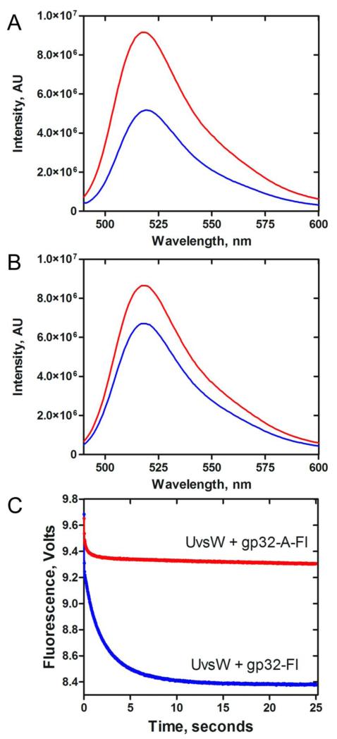
Fig 6. Interaction of gp32 and UvsW in the absence of DNA.
(A) Fluorescence of gp32-Fl in the presence (Blue) and absence (Red) of UvsW at an excitation wavelength of 480 nm. (B) Fluorescence of mutant gp32-A-Fl in the presence (Blue) and absence (Red) of UvsW excitated at wavelength of 480 nm. (C) Stopped flow kinetics of gp32-Fl binding to UvsW. gp32-Fl (500 nM) or gp32-A-Fl (500 nM) was rapidly mixed with UvsW (500 nM) and the reaction mixture was excited at 490 nm. The change in fluorescence signal was monitored using a 515 cut off filter. The resultant kinetic traces are shown in blue (gp32-Fl) and red (gp32-A-Fl).

