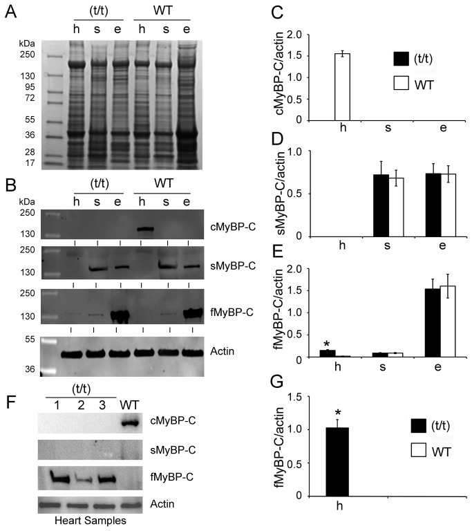
Figure 2. MyBP-C isoforms are differentially expressed in cardiac and skeletal tissue.
Ten µg of total proteins were resolved on 4-15% SDS-PAGE and stained with Coomassie Brilliant Blue (A). Expression levels of cardiac, slow skeletal and fast skeletal MyBP-C in (t/t) and WT mice were determined in heart (h), soleus (s) and EDL (e) muscles by Western blot analysis with respective antibodies (B). Representative Western blot shows that the presence of cardiac isoform of MyBP-C (cMyBP-C) is exclusive to ventricular muscle of WT mice (C), but completely absent in the (t/t) mouse hearts. sMyBP-C was significantly expressed both in the soleus muscle and EDL muscle (D). Conversely, fMyBP-C was mainly detected in EDL muscle (E), but significantly increased in the (t/t) hearts. All values were normalized to the expression of α-sarcomeric actin (MyBP-C/actin ratio) and expressed as relative values. The summarized quantitative data were derived from n=3 with mixed gender mice (*p< 0.01 versus WT). The increased level of fMyBP-C expression in the (t/t) hearts was reconfirmed by Western blot analysis (F) and not found in the WT hearts, summarized in panel G (n=6, *p< 0.0001 versus WT). α-sarcomeric actin was used as a loading control.

