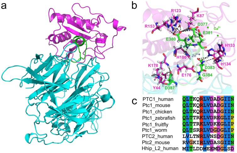
Figure 1. Complex structure of Shh-Hhip and sequence analysis of Hhip L2 with various species of Ptchs.
(a) The crystal structure of human Shh-Hhip complex (PDB ID: 3HO5). Shh, Hhip, and Hhip L2 (M373-D387) are represented in magenta, cyan, and green colors. The solvent molecules were omitted for clarity. (b) Hydrogen bond interactions between the Shh pseudo-active site (magenta) and Hhip L2 (green) are shown in black dotted lines and the hydrogen bonding residues are displayed as stick model. (c) The result of multiple sequence alignment between Hhip L2 and various species of Ptchs [21].

