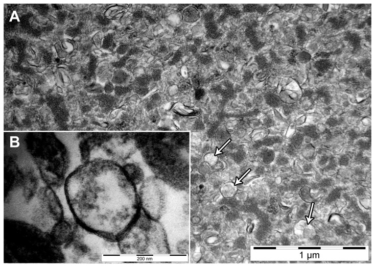
Figure 1. Transmission electron microscopy of mammary gland (MG) enriched plasma membrane vesicles (EPM).
A: Representative electron micrograph of EPM from lactating MG at 31’000 × magnification. Arrows depict single vesicles. B: The bilayer structure of the EPM from the same lactating MG at 230’000 × magnification. Electron micrographs of EPM isolated from non-lactating MG (not shown) were similar to that of lactating tissue.

