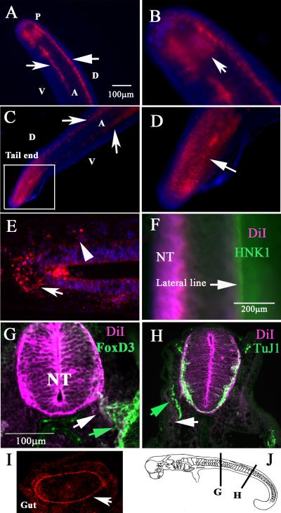
Figure 3. DiI labels migrating neural crest in Stage 26 & 29 C. punctatum embryos.
Shark embryo neural tubes were vitally labeled with DiI for 24hrs. Stage 26 shark embryo shows DiI cells as two bright lines (arrows in A) and as a sheet at tail end (arrow in B). Stage 29 shark embryo shows DiI cells as two bright lines (arrows in C) and as a sheet at the tail end (arrow in magnified area in D). Pseudo-longitudinal section showed delaminated DiI cells on top of the neural tube (arrow in E) and on one portion (rostral) of the somite (arrowhead in E). (F) shows whole-mount image of the embryo after HNK1 immunostaining of lateral line placodal neurons (arrow). (G) FoxD3 antibody stain (green arrow) co-localized with DiI cells (white arrow). (H) Anti-beta III tubulin labeling (TuJ1) neurons (green arrow) along the lateral line did not overlap with DiI cells (white arrow). (I) The developing gut had a line of DiI cells (arrow). Abbreviations: NT: neural tube; D: dorsal, V: ventral, A: anterior; P: posterior.

