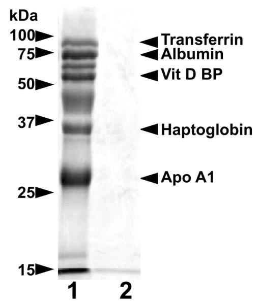
Figure 2. Selective proteinuria in BC with I-GS.
Shown are lanes from a silver-stained 15% SDS-PAGE gel loaded with urine proteins of an affected (lane 1) and a clinically normal BC (lane 2). Proteins in each lane were concentrated from urine samples containing 200 μg of creatinine. The affected dog was in metabolic remission at the time of urine collection due to previous parenteral cyanocobalamin administration. Identities of the labeled protein bands were confirmed by immunoblotting as previously reported [23-26].

