
Figure 3. Frameshift mutation near the CUBN exon/intron boundary 53.
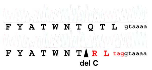
Shown are electophoretograms obtained by Sanger sequencing of exon 53 with flanking intronic sequence amplified by PCR from a normal (above) and an I-GS affected BC (below). The site of a single cytosine deletion in the affected BC sequence is indicated by the triangle. The deduced amino acid sequence is shown in single letter code below each electrophoretogram. The translation sequence and premature stop codon predicted by the frameshift are shown in red. The nucleotides indicated in lower case black are the splice donor sequence of intron 53.