
Figure 4. Deficient CUBN expression in ileum and kidney of BC with I-GS.
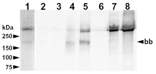
Shown is a blot of proteins accumulated from homogenates of ileal mucosa (lanes 1-5) and kidney cortex (lanes 6-8) of I-GS affected dogs (lanes 1-3 and 6) and normal controls (lanes 4, 5 and 7, 8). Lane 1 is an overexposure of lane 2 on the same blot. Twenty-five mg of total protein in ileal homogenates and 2 mg in kidney homogenates were incubated with 20 μL of IF-Cbl-agarose beads. Bound proteins were separated by non-reducing SDS-PAGE (4-20% gradient gel) and electroblotted. The membrane was incubated sequentially with rabbit polycolonal anti-canine CUBN serum [28] (1:20,000 dilution), goat anti-rabbit IgG-HRP conjugate, and chemiluminescent detection reagents. Migration of molecular weight markers is indicated on the left. The ileal brush-border size (~190 kDa) of CUBN that occurs after exposure of full-length CUBN to luminal proteases is indicated on the right (bb).