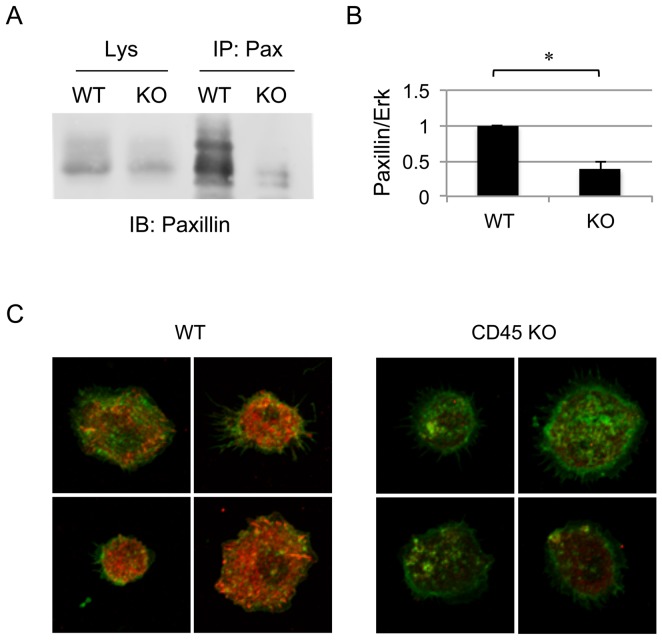
Figure 3. Paxillin expression is decreased in CD45 KO BMDM.
(A) Paxillin was immunoprecipitated from lysates of 107 day 7 WT or CD45 KO BMDM cells, followed by SDS-PAGE and Western blot with anti-paxillin. Paxillin levels were decreased in both immunoprecipitates and total lysates of CD45 KO BMDM compared to WT. Lysate control represents 4×10⁵ cell equivalents. (B) Quantification of paxillin in Western blots of BMDM lysates as represented by a ratio of paxillin in relation to the loading control (Erk). Represented is the average ratio obtained from five independent experiments. The asterisk indicates a difference with p<0.0001. (C) Immunofluorescence staining of paxillin (red) and actin (green) in day 7 CD45 KO and WT BMDM. Identical acquisition and analysis settings were used for all confocal images. Images are representative of two independent experiments.

