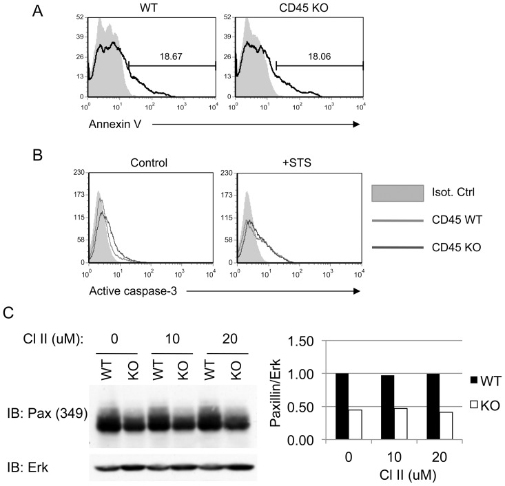
Figure 4. Inhibition of caspases does not restore paxillin expression in CD45 KO BMDM.
(A) Unstimulated day 7 WT or CD45 KO BMDM were stained with Annexin V (black line) or left unstained (grey, filled). Both cell types exhibited similar levels of basal Annexin V staining. (B) Day 7 WT or CD45 KO BMDM were treated with 5 µM of staurosporine (STS) for 4 hours (right panel), or DMSO control (left panel), and stained with anti-active caspase-3. (C) Day 7 BMDM were treated with the indicated concentration of caspase inhibitor II (CI-II) for 4 hours and lysed. Paxillin expression was assessed by Western blot. Anti-Erk was used as a loading control. Quantification of the density of the Western blot bands was done with Image J software. The image shown is representative of three independent experiments.

