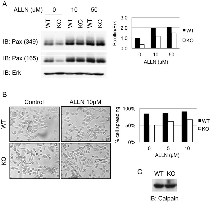
Figure 5. Inhibition of calpain restores paxillin expression and enhances cell spreading.
(A) Day 7 WT or CD45 KO BMDM cell lysates (106 cells) were treated with the indicated amounts of the calpain inhibitor ALLN for 4 hours. Lysates of 106 cells were run on SDS-PAGE gel and immunoblotted with two anti-paxillin monoclonal antibodies (clones 349 and 165) and anti-Erk as a loading control. The right panel shows the quantification of paxillin expression, probed with antibody from clone 349, relative to Erk was performed with ImageJ software. (B) Light microscopy image and quantification of cell spreading of WT and CD45 KO BMDM 4 hours after treatment with 10 µM of ALLN or DMSO (control) (C). All experiments shown are representative of three independent experiments.

