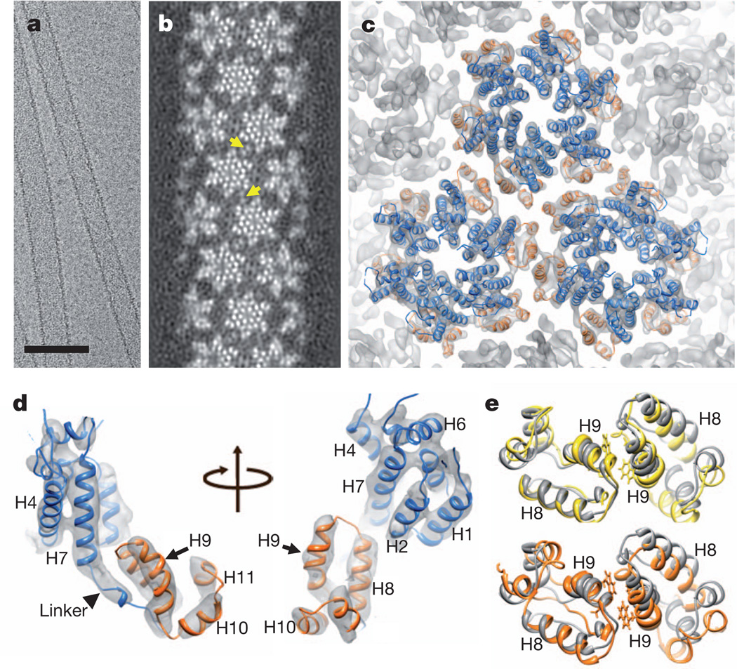
Figure 1. Cryo-EM reconstruction of HIV-1 CA tubular assembly at 8Å resolution andMDFF.
a, A cryo-EM image of recombinant A92E CA tubular assembly. Scale bar, 100 nm. b, Electron density map of the A92E CA tube with (−12, 11) helical symmetry. Yellow arrows indicate pairs of helix H9, located between adjacent hexamers. c, MDFF model of the HIV-1 capsid assembly, superimposed with the electron density map contoured at 4.0σ. Three CA hexamers, with NTDs (blue) and CTDs (orange), are shown. d,MDFF model of a CA monomer viewed from two angles. e, Two CTD dimer structures along -1 (orange) and 11 (yellow) helical directions, superimposed onto the NMR solution dimer structure (grey, 2KOD).

