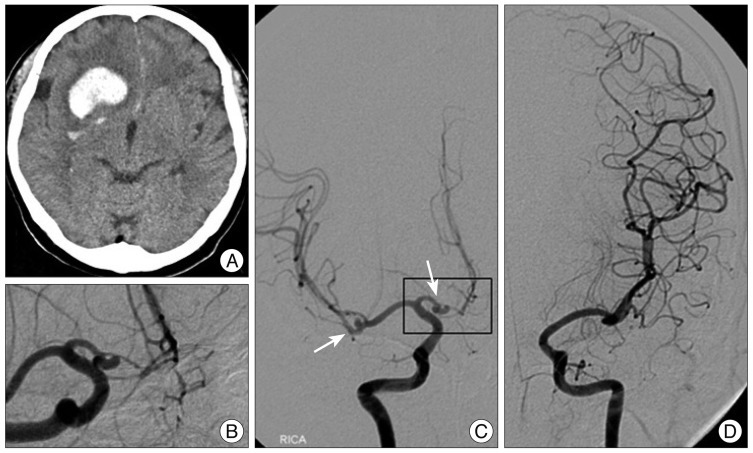
Fig. 1.
A : Computed tomography scan showing a large intracerebral hemorrhage in right side frontal lobe extending to basal ganglia. Subarachnoid hemorrhage was also identified at the anterior falx and sulci of medial frontal lobes. B : Angiography revealing an aneurysm arising from the proximal end of fenestrated right A1 segment. C : Right cerebral angiogram showing two aneurysms including one on the proximal end of fenestrated A1 and another on the middle cerebral artery bifurcation. Azygos anterior cerebral artery was visible on the angiogram. D : Left carotid angiogram showing that the right A1 segment is aplastic.

