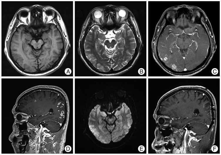
Fig. 1.
Radiologic findings. A and B : In brain MRI, there are low-signal intensity on T1-wighted axial images and high-signal intensities in the T2-weighed axial image on right occipital cortex. C and D : Multiple enhancing nodules with necrotic change are shown in right frontotemporoparietal cortex and white matter. E : In diffusion MRI, the high-signal intensity lesions are shown in right frontotemporoparietal cortex and white matter. F : The recurrence does not appear in the right temporal and occipital area with remaining focal enhanced lesions.

