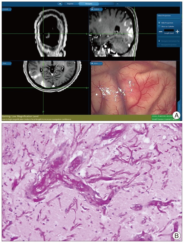
Fig. 2.

Operative and pathologic findings. A : There was no definitive mass lesion, but reddish tissues of the cortex and the thickened yellowish arachnoid membrane are shown. B : In the biopsy, there are shown the hyphaes with the positive periodic acid-shiff staining (original magnification ×200).
