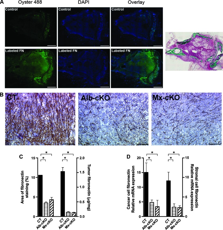
Figure 2.
The role of circulating fibronectin. (A) Labeled fibronectin can be detected within the tumor after injection. Masson-Goldner stain confirms the presence of cancer. Bars represent 500 µm. (B) Fibronectin staining was diminished in Alb- and Mx-cKO tumors. Bars represent 100 µm. (C) Quantifying the area of staining showed a decrease by more than 60% in Alb- and Mx-cKO tumors compared to controls (N = 3–4 mice/group). Similarly, quantifying the amount of fibronectin in tumors by ELISA reveals that CT tumors had more than 10-fold higher fibronectin content compared to Alb-cKO and Mx-cKO tumors. Fibronectin was examined by ELISA and values adjusted to protein (N = 3–6/group). (D) Both cancer cell fibronectin mRNA expression corrected to human HPRT and stromal fibronectin mRNA expression corrected to murine α-actin are decreased in Alb- and Mx-cKO (N = 4/group). *P < .05, **P < .001.

