Figure 5.
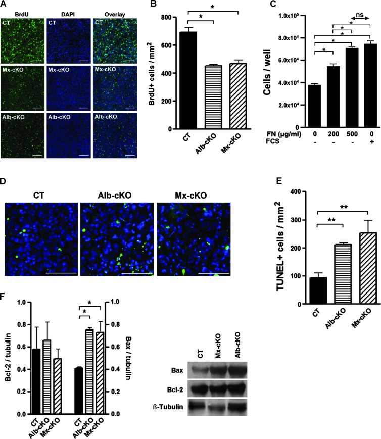
Effects of fibronectin content on proliferation and apoptosis. (A) The number of proliferating cells stained for BrdU in cKO tumors is diminished. Bars represent 100 µm. (B) Quantitative analysis of BrdU+ cells in tumors (N = 6 CT, 5 Alb-, and 5 Mx-cKO tumors). (C) Proliferation of tumor cells is improved by the addition of fibronectin. Fibronectin (500 µg/ml) and 10% fibronectin-depleted FCS increase proliferation to a similar extent. (D) TUNEL staining in tumor tissue from CT, Alb-, and Mx-cKO mice. Bars represent 200 µm. (E) The number of TUNEL-stained cells is increased in cKO tumors (N = 3–4/group). (F) Western blot analysis for Bcl-2 and BAX shows an increase in BAX in the absence of circulating fibronectin. A graph of densitometry of values adjusted to β-tubulin is shown on the left. On the right, an example is presented (N = 3/group).

