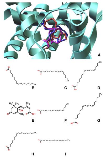
Fig. 2.
Chemical structure of naturally occuring agonists of PPARγ. (A) Representative binding mode of the most stable docked orientation of ligands with PPARγ illustrated in ribbon mode. Ligand poses are generated by AutoDock Vina. Cis-9, trans-11 CLA is in red; trans-10, cis-12 CLA is in magenta; PUA is in blue. (B) c-9,t-11 CLA. (C) t-10,c-12 CLA. (D) 2-D PUA. (E) ABA. (F) 2-CAA. (G) JAA. (H) α-ESA. (I) β-ESA. (J) Butyric acid, a type of SCFA.

