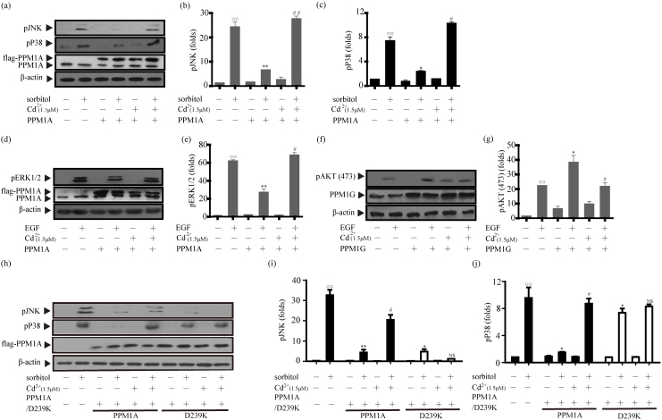
Figure 5. Cadmium suppresses PPM1A and PPM1G activity in cells.
HEK293 cells were incubated in DMEM supplemented with 10% FBS and transiently transfected with pcDNA3.1-flag-PPM1A or pcDNA3.1-flag-PPM1G. Sub-confluent HEK293 cells were serum-starved in DMEM for 12 h, incubated for 6 h with or without 1.5 μM CdCl2 and then treated with different stimulators. Cell lysate supernatants were prepared and analyzed by immunoblotting with specific antibodies. The data are from at least three experiments.  cells stimulated with EGF or sorbitol compared with unstimulated HEK293 cells;
  : P<0.05,
  : P<0.01. *: overexpression of PPM1A or PPM1G in HEK293 cells compared with control vectors; *: P< 0.05, **: P<0.01. #: Cells treated with CdCl2 compared with cells not treated with a metal ion; #: P< 0.05,##: P<0.01. (a and d) Cells were transfected with PPM1A or a control plasmid and then stimulated with sorbitol (0.4 M, 20 min, 37°C) or EGF (5 ng/ml, 5 min, 37°C) after incubation with or without 1.5 μM CdCl2. The levels of phosphorylated JNK, P38, and ERK were monitored by immunoblotting with specific antibodies. Actin was used as a loading control. (b, c and e) Statistical analysis of the data presented in panels a and d. (f) Cells were transfected with wild-type PPM1G and stimulated with 5 ng/ml EGF for 5 min at 37°C after incubation with or without 1.5 μM CdCl2 for 6 h. The level of phosphorylated AKT(473) was monitored by immunoblotting with specific antibodies. (g) Statistical analysis of the data in panel f. (h) Cells were transfected with the control, PPM1A, or PPM1A D239K plasmid and then stimulated with sorbitol (0.4 M, 20 min, 37°C) after incubation with or without 1.5 μM CdCl2. The levels of phosphorylated JNK, P38 were monitored. (i and j) Statistical analysis of the data in panel h.

