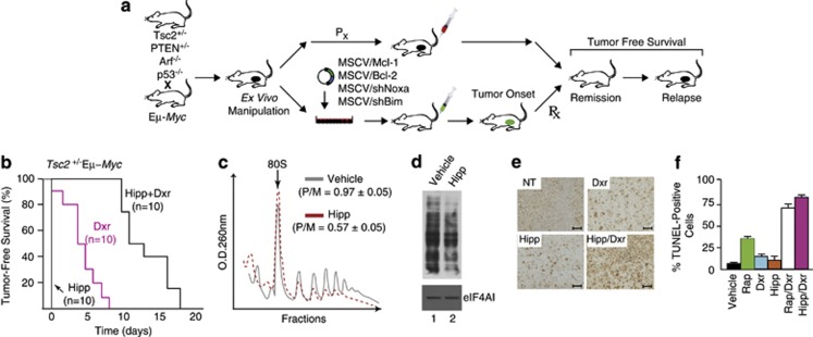
Figure 1.
Hipp alters chemoresistance to DNA damaging agents in Eμ-Myc lymphomas. (a) Schematic illustrating generation of Eμ-Myc tumors of defined genotypes. Indicated are the ex vivo and in vivo manipulations used in this study to assess tumor growth and drug response. (b) Kaplan–Meier plot demonstrating response of Tsc2+/−Eμ-Myc tumor-bearing mice to Hipp, Dxr, and Hipp+Dxr treatment. P<0.001 for Hipp+Dxr versus Dxr. (c) Hipp inhibits translation in Tsc2+/−Eμ-Myc tumors in vivo. Mice bearing Tsc2+/−Eμ-Myc tumors were treated with vehicle or Hipp (10 mg/kg) and 4 h later, tumors were excised, cytoplasmic extracts prepared and polysomes resolved. The position of 80S ribosomes is indicated and the polysome/monosome (P/M) ratios from three independent experiments denoted. (d) Hipp inhibits translation in vivo. Mice bearing Tsc2+/−Eμ-Myc tumors were treated with vehicle or Hipp (10 mg/kg) and 4 h later were injected with 100 mg/kg puromycin. One hour later, tumors were excised, cytoplasmic extracts prepared, proteins resolved by SDS-PAGE and transferred to Immobilon-P by western blotting. Blots were probed with an anti-puromycin (upper panel) or an anti-eIF4AI (lower panel; loading control) antibody. (e) Representative micrographs of Tsc2+/–Eμ-Myc lymphoma sections stained by TUNEL assay; bars represent 50 μm. C57BL/6 mice bearing well-palpable tumors were administered vehicle or Hipp. Twenty-four hours later, mice received Hipp, Dxr, or a combination of Hipp+Dxr. Six hours after treatment, tumors were extracted and stained. (f) Quantification of tumor cells positive for TUNEL staining following treatments described in Panel E and Suppl Figure 1. The cell count was obtained from two different fields taken from two sections (n=4). Results are expressed as the percent mean±s.d.

