Figure 1.
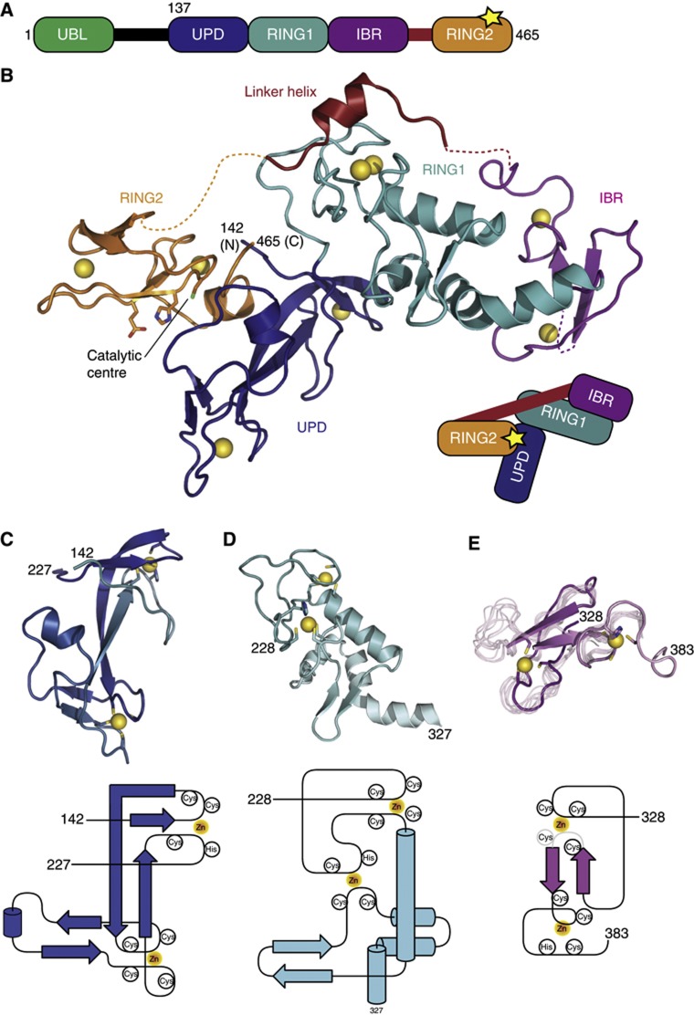
Structure of Parkin. (A) Domain structure of Parkin. A yellow asterisk (*) indicates the catalytic Cys431 in RING2. (B) Structure of the Parkin UPD-RBR domain. Individual domains are coloured blue (UPD), cyan (RING1), purple (IBR) and orange (RING2). The linker helix is shown in red and the Zn atoms as yellow spheres. Putative catalytic residues in RING2 are labelled. Dotted lines indicate disordered stretches. Terminal residue numbers are indicated. A cartoon based on (A) depicts domain interactions. (C) Structure and topology of the UPD. Residues coordinating Zn atoms are shown. (D) Structure and topology of the extended RING1. (E) Structure of the crystallized IBR domain with eight superposed models from the previously described NMR ensemble (pdb-id 2jmo, Beasley et al, 2007). The unresolved loop in Parkin IBR is most flexible also in NMR analysis. The topology is shown below, the region in grey is disordered in the structure.

