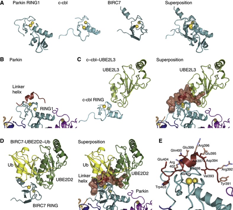
Figure 4.
RING1–E2 interactions. (A) Comparison of Parkin RING1 with RING domains of c-cbl (pdb-id 1fbv, Zheng et al, 2000) and BIRC7 (pdb-id 4auq, Dou et al, 2012). Zn atoms are shown as yellow spheres. (B) Parkin RING1 in context of the RBR, with linker helix bound. (C) Left—structure of c-cbl RING domain bound to UBE2L3, right—superposition of Parkin RING1 and c-cbl RING. The linker helix is shown under a semitransparent surface. (D) Representation as in (C) for BIRC7 bound to UBE2D2∼Ub. The Parkin linker helix blocks E2 and Ub interactions. (E) Detail of linker helix:RING1 interactions. Transparent side chains were disordered in the crystal structure and were included in their favoured rotamer to indicate the amphipathic nature of the helix.

