
Fig. 2.
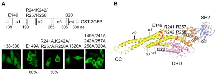
Identification of residues essential for the STAT5a NLS. (A) Location of alanine substitutions in the STAT5a coiled-coil domain. Below are fluorescence images of STAT5a coiled-coil domain tagged with GST-2GFP with the single or combined mutations noted above the image. Images represent the entire population of cells in culture, unless noted as 80% for E149A and 30% for 241A, 242A/257A and 258A. (B) Position of mutated residues in a ribbon diagram of the crystal structure of STAT5a coiled-coil domain (Protein Data Bank ID code 1Y1U). E149A in the first α-helix (α1), basic residues (R241A, K242A/R257A, R258A) in the second α-helix (α2) and I320A in the fourth α-helix (α4) are indicated.