Fig. 2.
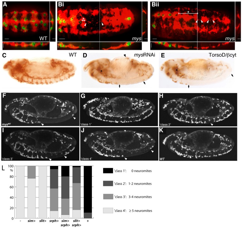
Environmental and haemocyte specific requirements for βPS integrin. (A,B) Orthogonal projections of ventrally orientated stage 15 WT and mys mutant embryos with haemocyte-specific expression of GFP (green), injected with dextran dye (red) to reveal spatial constraints surrounding the haemocytes. Scale bars: 20 µm (ventral view); 5 µm (orthogonal projections). (A) Within injected WT embryos the dye permeates along the length of the embryo, indicating VNC–epithelial separation. (B) Within mys mutant embryos spreading of the dye becomes restricted, indicating incomplete VNC–epithelial separation (white arrowheads indicate restricted area of ventral midline). (i) In some embryos the absence of dye coincides with the distance reached by the lead haemocyte migrating from the anterior of the embryo along the ventral midline, whereas in others (ii) the lead haemocyte fails to reach the area where the dye becomes restricted (distance between lead haemocyte and spatial restriction indicated by line and asterisk), suggesting that spatial constraint is not the sole cause of disruption to haemocyte migration along the ventral midline. (C–E) Lateral view of fixed stage 13 embryos. Haemocyte myospheroid requirements were assessed by coexpressing UAS-CD2 (C) and either UAS-mysRNAi (D) or a dominant-negative version of the mys subunit, UAS-TorsoD/βcyt (E), under the control of the srph-GAL4 driver, and staining with an anti-CD2 antibody. Expression of either UAS-mysRNAi or UAS-TorsoD/βcyt phenocopies the haemocyte migration defects observed in mys mutant embryos. (F–K) Lateral views of fixed and stained stage 13 embryos. (F) mysM/Z embryo; (G–J) grading of embryos into ‘classes’ based on level of haemocyte migration rescue; and (K) WT embryo. (L) Quantification of haemocyte migration rescue at stage 13 when UAS-mys is expressed in the midline under the control of the sim and slit GAL4 drivers, and/or in the haemocytes under the control of srph-GAL4.

