
Fig. 4.
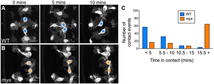
myospheroid plays a role in contact repulsion. (A,B) Stills taken from live-cell imaging of GFP-expressing haemocytes in WT and mys mutant embryos undergoing random migration at stage 15. (A) In WT embryos contacting haemocytes (blue asterisk) demonstrate contact repulsion, rapidly repolarising and migrating away from one another. (B) In mys mutant embryos haemocytes remain in contact, unable to undergo contact repulsion (orange asterisks). Scale bars: 10 µm. (C) Quantification of the time the lamellipodia of two haemocytes remain in contact. There is a dramatic increase in this time interval in mys mutant embryos (average time in contact for WT and mys haemocytes was 5.6 and 27.1 minutes, respectively), P>0.01, n = 89 (WT) and n = 43 (mys) contact events.