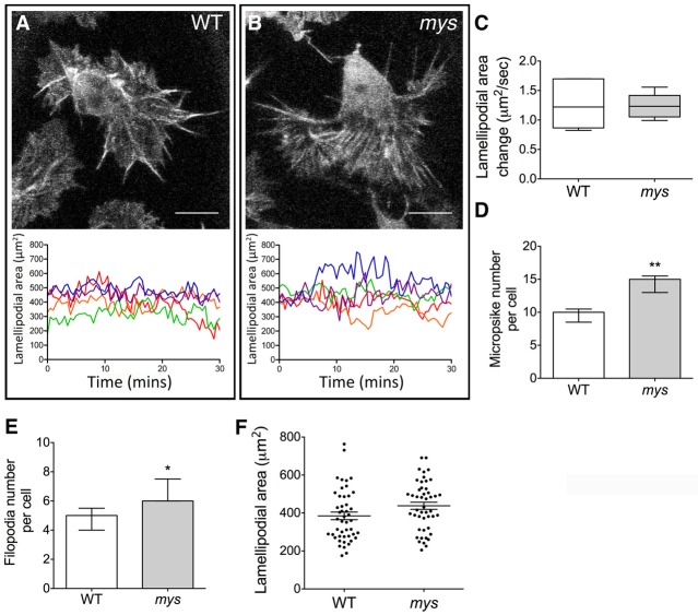
Fig. 6.
Haemocytes lacking myospheroid show altered actin dynamics. (A,B) Stills taken from live-cell imaging of haemocytes expressing LifeAct under the control of srp-GAL4 in WT and mys mutant embryos. Scale bars: 10 µm. The graphs beneath show the lamellipodial area of five haemocytes measured at 30 second intervals over a 30 minute time period. The large fluctuations in WT and mys mutant haemocytes indicate that the overall lamellipodial dynamics remain unchanged in the absence of integrin βPS. (C) This was confirmed when the average lamellipodial area change per haemocyte is compared with that in WT (n = 5 haemocytes per genotype). (D,E) Other actin-dependent structures within the haemocytes are affected in the mys mutant, with (D) an increase in the number of microspikes compared to WT (average 4.8 and 6.6, respectively, P<0.05) and (E) the number of filopodia per haemocyte (average 9.6 and 14.4, respectively, P<0.05). (F) Quantification reveals no difference in the lamellipodial area of WT and mys mutant haemocytes (n = 47 haemocytes for both genotypes).

