Fig. 2.
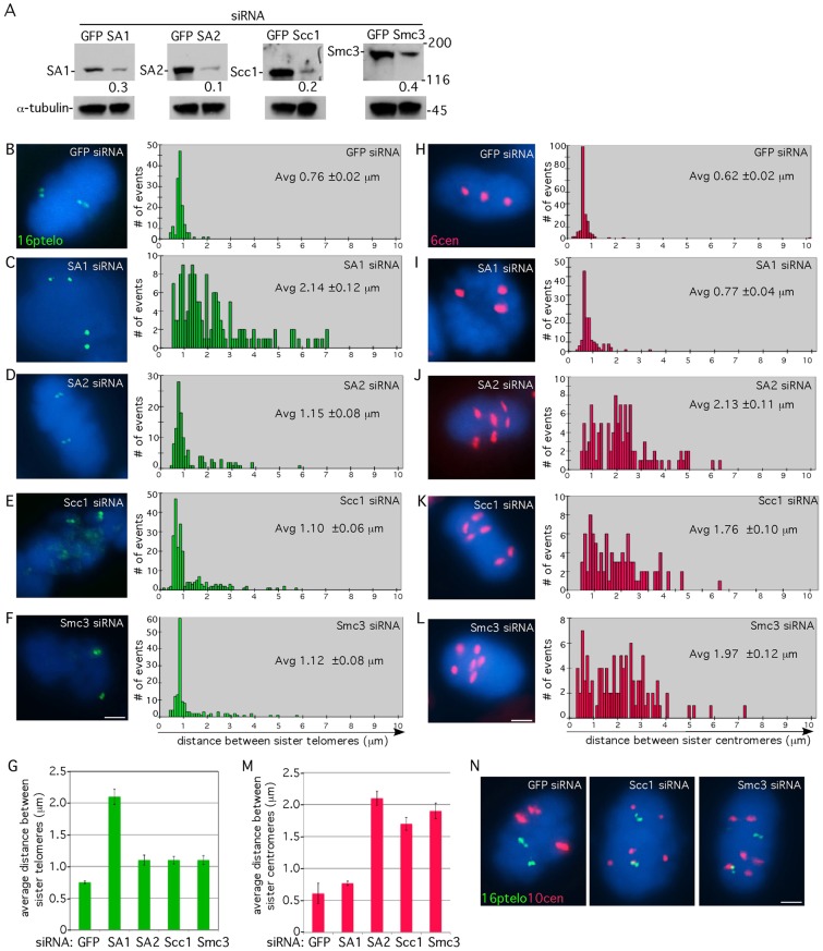
Telomere cohesion is maintained in mitosis in cohesin-ring-depleted cells. (A) Immunoblot analysis of extracts from HeLaI.2.11 cells transfected with siRNA to GFP, SA1, SA2, Scc1 or Smc3 for 48 hours and probed with the indicated antibodies. Protein levels relative to α-tubulin and normalized to the GFP siRNA control are indicated below the blots. (B–F) Telomere and (H–L) centromere FISH analysis of HeLaI.2.11 cells isolated by mitotic shake-off following 48 hours transfection with GFP (B,H), SA1 (C,I), SA2 (D,J), Scc1 (E,K) or Smc3 (F,L) siRNA with a 16ptelo (green) or 6cen (red) probe. The cen locus is trisomic. DNA was stained with DAPI (blue). Scale bars: 5 µm. Histograms showing the distance between FISH signals (n = 98–225) are on the right with the average (Avg) distance (± s.e.m.) indicated. (G,M) Graphical representation of the average distance (± s.e.m.) between sister telomeres (G) or centromeres (M). (N) Combined telomere and centromere FISH analysis. HeLaI.2.11 cells were transfected with GFP, Scc1 or Smc3 siRNA for 48 hours and probed with 16ptelo (green) and 10cen (red). The cen locus is trisomic. DNA was stained with DAPI (blue). Scale bar: 5 µm.

