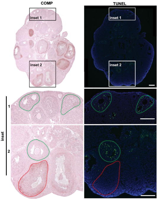
Figure 6.
Follicles that express COMP are TUNEL-negative. Ovarian section from a prepubertal mouse 48 hr after injection with eCG immunohistochemically stained for COMP. Adjacent ovarian sections stained for TUNEL. Follicles delineated in green and red dashed lines highlight COMP-positive and TUNEL-positive staining, respectively. Scale bars, 200 μm. Images are representative of three independent experiments.

