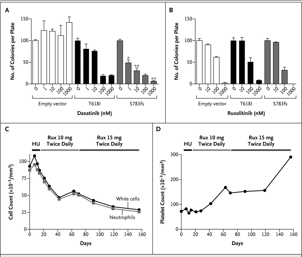
Figure 2. Use of Tyrosine Kinase Inhibitors to Treat Dysregulated Signaling Induced by CSF3R Mutations.
Panel A shows the effect of dasatinib on colony formation in bone marrow cells from mice that were infected with mutant CSF3R-containing retroviruses or an empty vector; the control cells expressed endogenous wild-type CSF3R. Cells were grown in methylcellulose containing the minimal amount of granulocyte colony-stimulating factor (G-CSF) necessary to form colonies (10 ng per milliliter for the empty vector, 0.4 ng per milliliter for the S783fs mutation, and no G-CSF for the T618I mutation). Cells were plated with increasing concentrations of dasatinib (0, 1, 10, 100, and 1000 nM). The experiment was performed in triplicate with the number of colonies normalized to those in the untreated controls. Values represent the mean percent colonies; the T bars indicate standard errors. A single asterisk indicates P<0.07, and a double asterisk indicates P<0.005 for the comparison between the T618I mutation and the S783fs mutation at equivalent doses of dasatinib. Panel B shows the results of a similar colony-formation assay, in which the cells were plated with ruxolitinib (0, 10, 100, or 1000 nM). Panel C shows the results for Patient 9, who had CNL and a CSF3R T618I mutation and in whom earlier testing indicated sensitivity to ruxolitinib (Rux) in vitro (Fig. S3C and S3E in the Supplementary Appendix). This patient was treated with 500 mg of hydroxyurea (HU) daily starting on day 13. Hydroxyurea was stopped on day 21 and oral ruxolitinib (at a dose of 10 mg twice daily) was administered. On day 70, the dose of ruxolitinib was increased to 15 mg twice daily. The numbers of white cells and neutrophils (absolute neutrophil count) are shown. Panel D shows normalized platelet counts while Patient 9 was undergoing the treatment regimen shown in Panel C.

