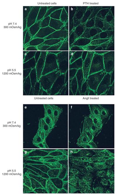
Figure 6. AT1R receptors internalize following AngII stimulation under `harsh' inner medullary conditions, whereas PTH-stimulated PTHR does not.
LLC-PTHR-GFP (a–d) and LLC-AT1R-GFP (e–h) cells, stably expressing either PTHR-GFP or AT1R-GFP were incubated in the absence (a, c and e, g) or presence of 1 μm of their respective agonists (b, d and f, h). Cells were incubated with either neutral (a, b and e, f) or acidic hyperosmotic medium (c, d and g, h). Sequential images were acquired using spinning disk confocal microscopy. Acquired images of cells taken before and after 30 min of agonist stimulation are representative of four independent experiments.

