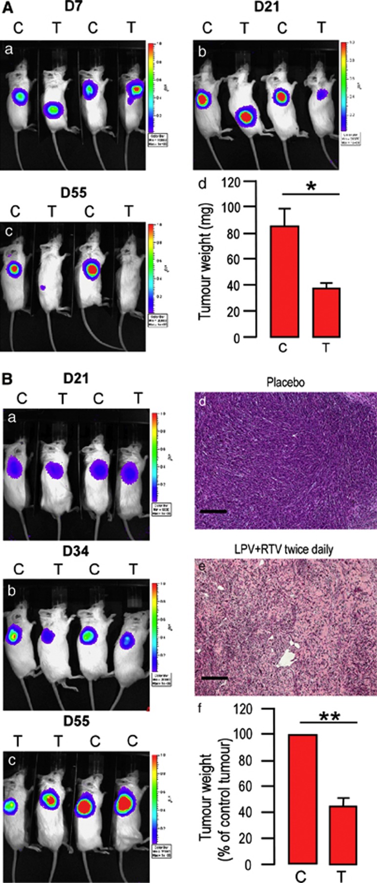
Figure 5.
Fixed association of LPV/RTV decreases CSCs-induced allograft formation. CB17/SCID mice transplanted with 250 000 cells from an adenocarcinoma (A) or an intestinal tumor (B) were treated twice daily with placebo or LPV/RTV. In vivo bioluminescent imaging of light emitted by cells reveals a decrease in the size of sites for light emission in mice receiving LPV/RTV after 21 days (Panel A, b) or 34 days (Panel B, b) of treatment, and this was more pronounced after 55 days of treatment. Tumor weight was assessed after 55 days of treatment and was significantly lower in mice receiving LPV/RTV as compared with those receiving placebo (panel A, d; panel B, f) (n=5, *P<0.05; **P<0.01). Histological analysis of the allografts shown in panel B (d–e) confirms that LPV/RTV impaired cell proliferation and allograft formation (scale bar, 100 μm)

