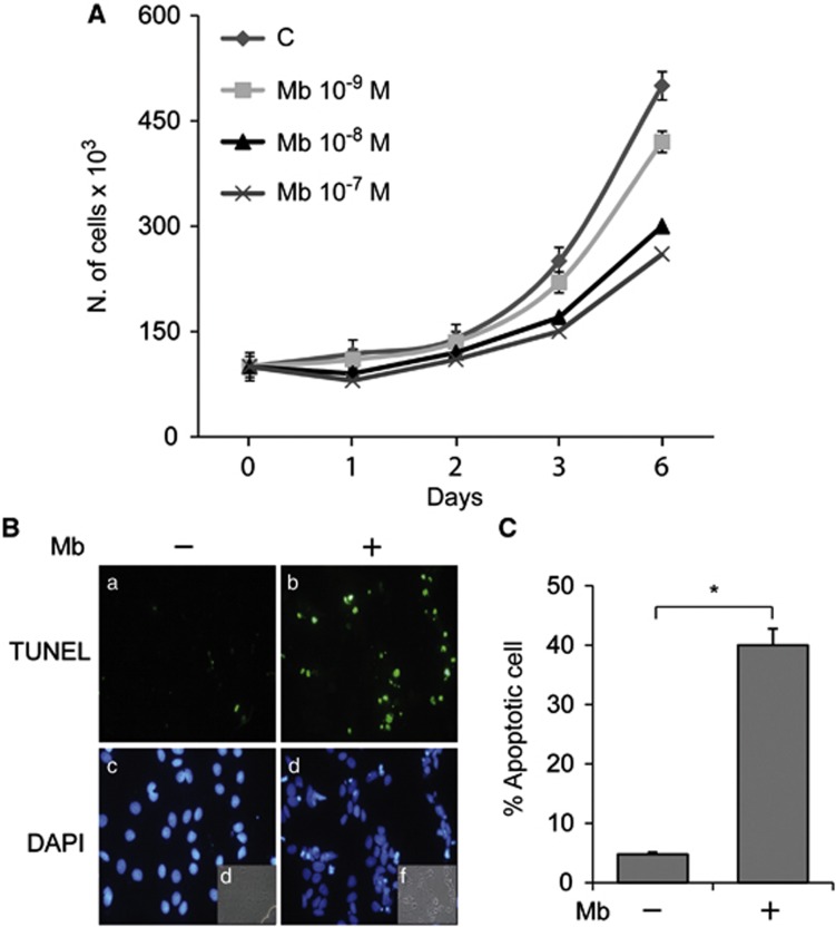
Figure 1.
Long-term Mb administration inhibits MCF-7 cell proliferation and induces apoptosis. (A) Cell counting by trypan blue exclusion test in cells treated as indicated (6 days). (B) Cells treated with 10−8 M Mb for 6 days were subjected to TUNEL nuclear staining (a, b) and viewed by a fluorescent microscope. DAPI staining for nuclei detection (c, d); bright field (e, f). Apoptotic cells were photographed at × 10 magnification and then counted using Image J software. (C) Histograms represent the apoptotic index % apoptotic cells/total cell number in the field). *P<0.01

