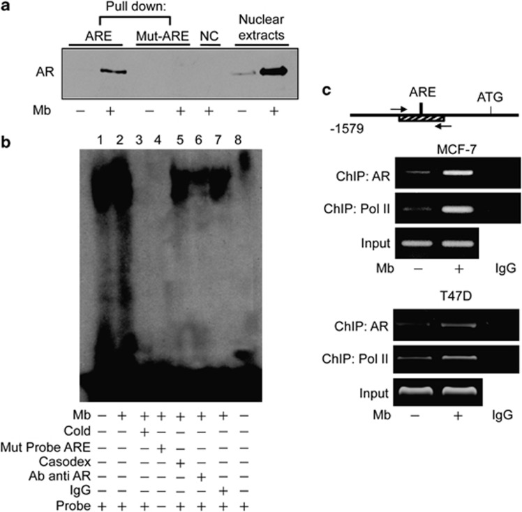
Figure 5.
Ligand-activated AR binds to an ARE site within DAX-1 promoter. (a) DAPA on nuclear extracts from MCF-7 cells treated with 10−8 M Mb for 2 h. Wild-type (ARE) or mutated (mut-ARE) biotinylated oligonucleotide were used. Unbound fraction, negative control (NC); nuclear extracts, positive control. (b) EMSA on nuclear extracts from MCF-7 cells untreated (lane 1) or treated with 10−8M Mb for 2 h (lane 2-7). 100-fold molar excess of unlabeled probe (lane 3); labeled mutated probe (lane 4); addition of 10−5M Casodex (lane 5); pre-incubation with anti-AR antibody (lane 6) or IgG (lane 7); probe alone (lane 8). (c) Sheared chromatin from MCF-7 or T47D cells treated with 10−8M Mb for 2 h was precipitated using anti-AR or anti-RNA Pol II antibodies. IgG, control samples. DNA input, loading control. Results are representative of three independent experiments

