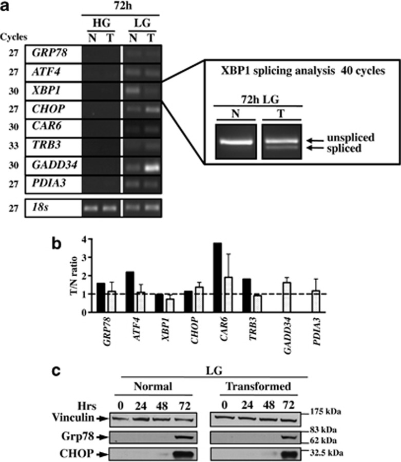
Figure 3.
Semi-quantitative RT-PCR and western blot analysis indicated that UPR is activated at LG. (a) Semi-quantitative RT-PCR of the mRNAs specific for different UPR-related genes in normal (N) and transformed cells (T) at 72 h in HG and LG. (b) Comparison between the relative levels of expression calculated by Affymetrix and semi-quantitative RT-PCR for the same genes at LG. (c) Western blot analysis of UPR activation upon glucose depletion. To follow UPR activation, the expression of Grp78 and CHOP proteins was analyzed. Images are representative of at least three independent experiments

