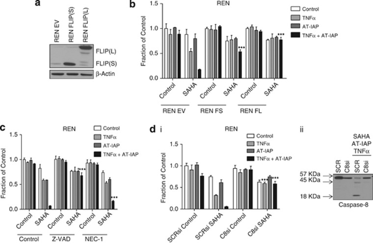
Figure 7.
(a) Western blot analysis of FLIP expressing in control (EV) REN cells and cells stably overexpressing FLIP(S) (FS) and FLIP(L) (FL). (b) Cell viability assay of REN EV, FS and FL overexpressing cell lines pretreated with 2.5 μM SAHA for 12 h before treatment with 10 μM AT-IAP, 10 ng/ml TNFα or a combination of AT-IAP and TNFα for 48 h. (c) Cell viability assay of REN cells pretreated with 2.5 μM SAHA for 12 h before treatment with 10 μM AT-IAP, 10 ng/ml TNFα or a combination of AT-IAP and TNFα for 48 h. Cells were incubated with 10 μM z-VAD-fmk (Z-VAD) or 30 μM necrostatin-1 (NEC-1) for 1 h before treatment with AT-IAP and TNFα. (d) (i) Cell viability assay of REN cells transfected with SCRsi or caspase 8 (C8si). Cells were pretreated with 2.5 μM SAHA for 12 h before treatment with 10 μM AT-IAP, 10 ng/ml TNFα or a combination of AT-IAP and TNFα for 48. (ii) Western blot analysis of caspase 8 expression in REN cells transfected with SCRsi or caspase 8 (C8si) siRNA. Cells were pretreated with 2.5 μM SAHA for 12 h before treatment with 10 μM AT-IAP and 10 ng/ml TNFα combination for 48 h

